multi-modual oct
The PTS 2000 is our top of the line Automated Projection Perimeter, with the latest in visual field testing standards. Utilizing the most advanced testing techniques available, the PTS 2000 provides fast testing strategies, customizable patterns, kinetic testing and all Goldmann stimuli sizes.
The PTS 925Wi is our small but powerful Automated Perimeter, with more features than you would expect. This unit shares most of the same great features of the PTS 2000. Unlike most space-saving perimeters, the PTS 925Wi allows peripheral testing outside the central 30 degrees.
Capture 10 additional central field locations with the new 24-2C pattern.
Let our voice guide provide instructions for the patient during an exam. In 12 different languages.
Switch between dark and light view. Reduce eye strain during testing in a darkened room.
Perform automated or semi-automated Kinetic perimetry with the PTS 2000.
Using ZETA™ thresholding algorithms, test your patients in under 3 minutes.
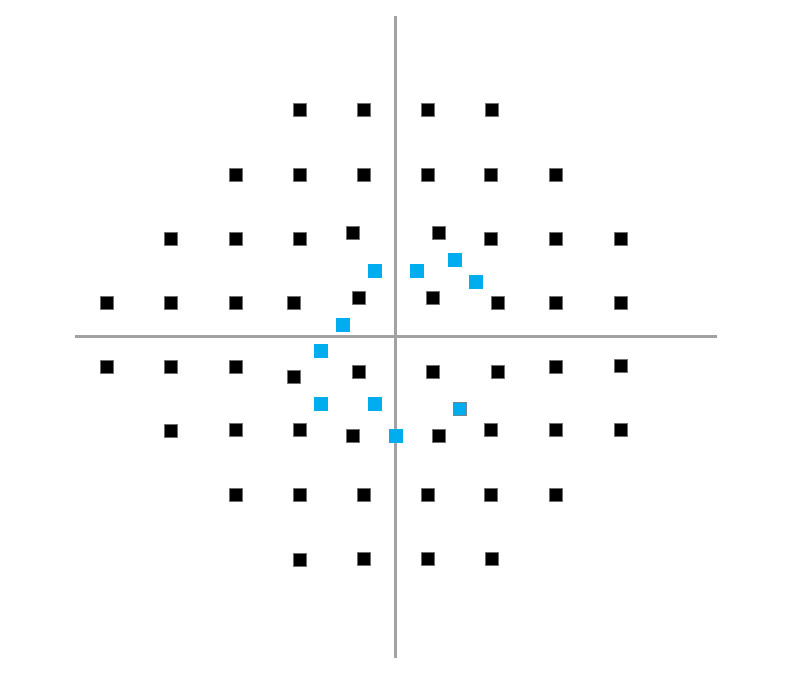
Monitor patient pupil movement during examination.
Our software detects when the eye is closed, and will not present stimuli to the patient.
Live monitoring and automated adjustments throughout exams.
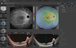
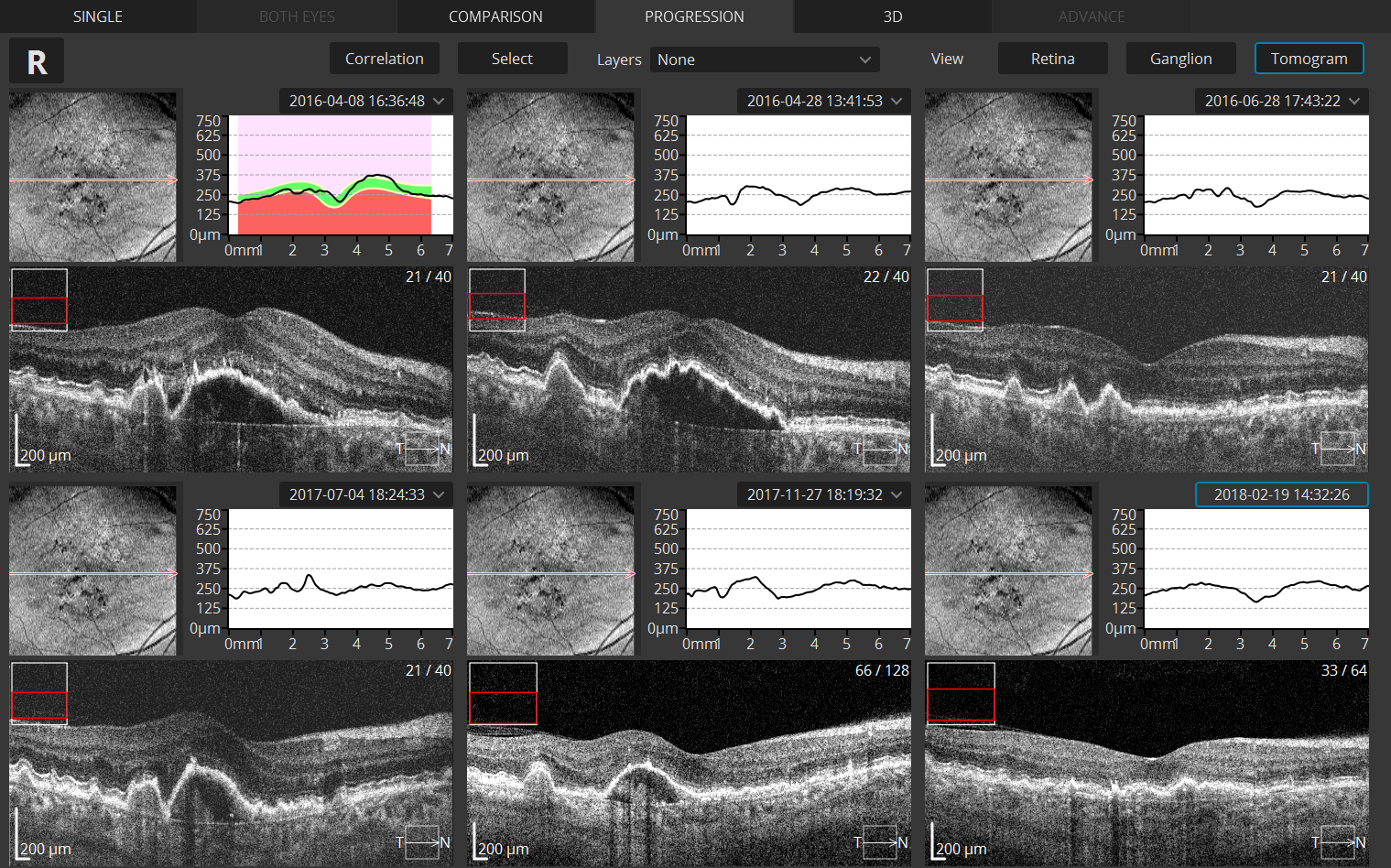
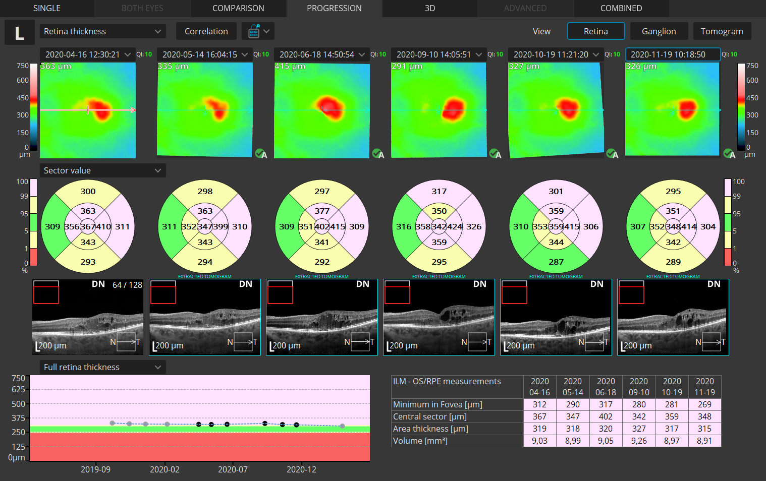
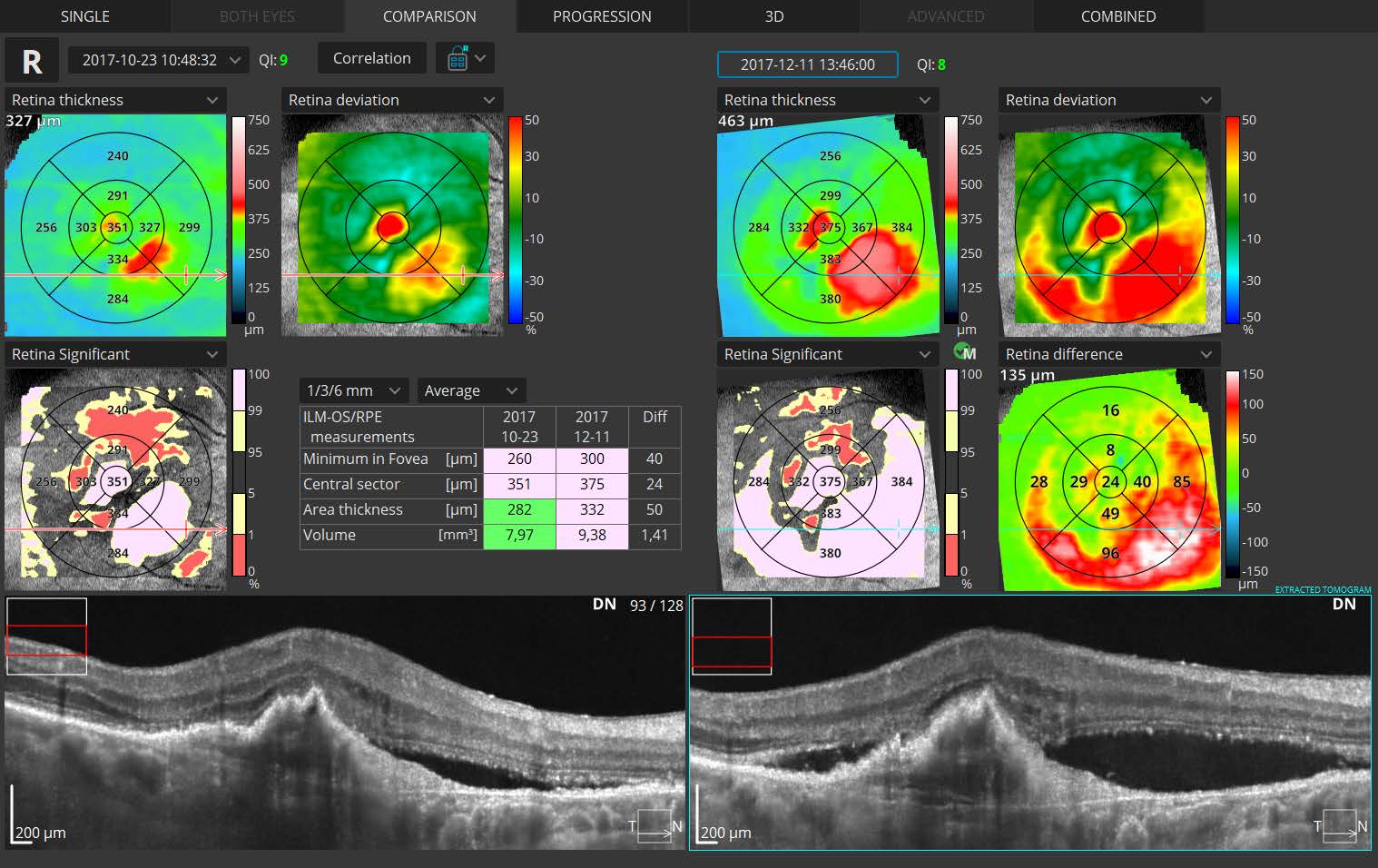
Create a baseline and follow disease progression over time.
In the review screen, click on any point during the exam to see the virtual eye position.
Voice messages assist operator and patient during the examination.
BluGlow™ light band provides a minimal ambient light to the darkened testing area, providing operator and patient with the ability to navigate their surroundings.
Both our perimeters use the same great software. Perfect for seamless training across satellite offices.
How can we beat the prices of our competitors?
Buy Optopol and save. You get a full suite of diagnostic tools for a great price.
We will never ask you to purchase a service contract. Our industry leading warranty keeps you covered.
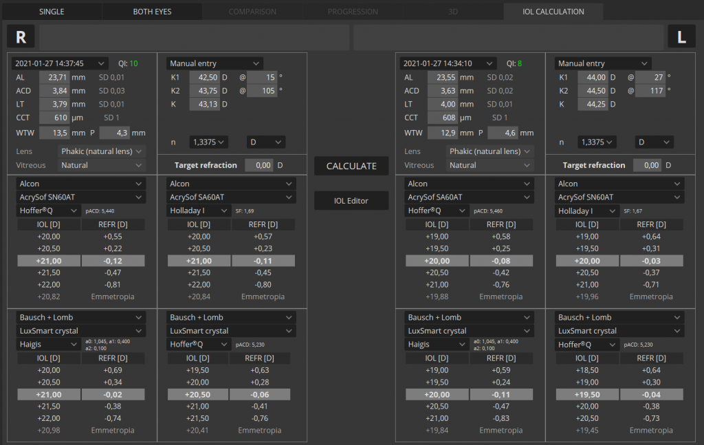
Humphrey and Octopus style included. View your exams the way you want, set default or change on-the-fly.
Import your old HFA data. Browse all your patients and exams through our software.
Windows 10 Pro and review software included. Our devices easily network to your EMR and DICOM system.
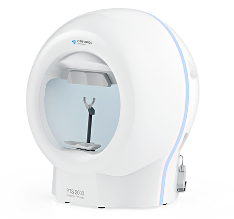
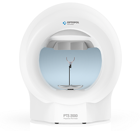
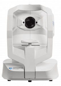
This is our 2nd generation Perimeter. A powerful 4" x 4" mini PC runs our software and can be easily upgraded or swapped out.
multi-modual oct
iTracking™ technology compensates for involuntary eye movements and blinks.
When OCT scan is used, each anatomical region is acquired twice automatically. The system immediately creates an artifact free MC examination using the Motion Correction TechnologyTM.
The elimination of eye movement and blinking artifacts ensures the highest resolution of Angio OCT images without patient inconvenience. Clear OCTA data set allows easier interpret retina vasculature condition.
multi-modual oct
RETINA
A single 3D Retina examination is sufficient to perform both Retina and Glaucoma analysis based on retinal scans. During the analysis the software automatically recognizes 8 retina layers to ensure a more precise diagnosis and mapping of any changes in the patient’s retina condition.
High density of standard 3D scan allows the operator to precisely track the disease progression. The operator can analyze changes in morphology, quantified progression maps and evaluate the progression trends
multi-modual oct
The software can track 3D scans and register them to the OCT baseline exam by recognizing patterns in the shape of blood vessels. Active tracking and post-processing point-to-point registration allows the user to precisely see and track the changes in retina morphology in Comparison and Progression analysis.
This module allows visualization of the retinal microvasculature. Angiography SOCT is a non-invasive, dye-free technique providing 3D image of retinal blood circulation.
The quantification tool provides quantification of the vasculature in the whole analyzed area together with values in specific zones and sectors. Thanks to the heat map of the analyzed vasculature the evaluation of vascular structure conditions is much faster. The choice of the quantification method increases the sensitivity of analyses for specific deseases.
FAZ – Foveal Avascular Zone measurements enable the quantification and monitoring of changes in Superficial and Deep vascular layers. The FAZ tool is also available for narrow and wide scans.
NFA – Non Flow Area measurement tool makes it possible to quantify the Non Flow Area on the OCT Angio examination. It provides the sum of all marked areas.
VFA – Vascular Flow Area allows the user to examine the pathologically affected area and to precisely measure the area covered by vascularization.
The simple and easy area measurement can be performed on a predefined or user-selected vascular layer.
Comprehensive glaucoma analytical tools for quantification of the Nerve Fiber Layer, Ganglion layer and Optic Head with DDLS enable the user to perform precise diagnosis and monitoring of glaucoma over time.
With the golden standard 14 optic nerve parameters and a new Rim to Disc and Rim Absence the description of ONH condition is quick and precise.
Advanced view which provides combined information from Retina and Disc scan to integrate details of the Ganglion cells, RNFL, ONH in a wide field perspective for comprehensive analysis.
Asymmetry analysis of Ganglion layers between hemispheres and between eyes helps to detect and identify glaucoma in early stages and in non-typical patients.
To eliminate the common problem of understanding the patient’s IOP, the pachymetry module provides IOP Correction value. With
the implemented Adjusted IOP formula, you can quickly and precisely understand the measured IOP value. The Pachymetry and
Anterior Chamber Angle Verification require no additional attachments. The predefined Glaucoma protocol, which consists of Retina,
Disc and Anterior scans, can be done automatically to reduce patient chair time.
* Images curtesy of Edward Wylęgała, MD, PhD
Comprehensive Glaucoma Solution
Combined OCT and VF results analysis
Comprehensive structure & function report includes the following:
Built-in anterior lens allows the user to perform the imaging of the anterior segment without installing additional lens or forehead adapter. Now you can display the whole anterior segment or focus on a small area to bring out the details of the image.
Anterior Chamber exam with a fast view of the whole Anterior Chamber make the evaluation of gonioscopy situation and the verification of cataract lens easier and faster.
See incredible details of a selected area of the anterior segment.
A proficient networking solution increases productivity and enhances patient experience. It allows you to view and manage multiple examinations from review stations in your practice. It also effortlessly facilitates patient education by allowing you to interactively show examination results to patients. Every practice will have different requirements which we can cater for by tailoring a bespoke service. There is no additional charge for the server module.
Store, exchange, and transmit results through DICOM gateway to the hospital network.
This optional software module allows visualization of the retinal microvasculature. Angiography SOCT is a non-invasive, dye-free technique providing 3D image of retinal blood circulation.
Software allows to observe, track and compare changes in the microvasculature of the retina in both eyes.
Available quantification methods:
Quantification is available for a specific layer in Angio OCT exam:
The standard version of the module broadens the functionality of the device with the addition of the IOL Tab which allows the user to calculate the lens power of a selected IOL model (on the basis of the patient’s morphological data).
The basic B-OCT® module provides an exceptional comfort of daily use.
The Angiography mosaic delivers high-detailed images over large field of the retina.
Advanced tab provides: view of any vascular layers, enface view of vascular layers, depth coded and thickness map. Mosaic modes: 10×6 mm, 12×5 mm, 7×7 mm, 10×10 mm and Manual (up to 12 images).
IOL formulas allow the user to calculate IOL implant parameters. Our systems now support the latest IOL data base standard IOLCon.org so that you can always keep your library up-to-date.
T-OCT™ is a pioneering way to provide detailed corneal Curvature maps by using posterior dedicated OCT. Ante-rior, Posterior surface and Corneal Thickness allow to provide the True Net Curvature information. With Net power, the precise understading of the patient’s corneal condition comes easily and is free of errors associated with modelling of posterior surface of the cornea. SOCT T-OCT module provides Axial maps, Tangential maps, Total Power map, Height maps, Epithelium and Corneal thickness maps.
Corneal topography module clearly shows the changes in the cornea on the difference map view. Customize your
Topography module provides:
Easily detect and classify keratoconus with the Keratoconus classifier. Classification based on KPI, SAI, DSI, OSI and CSI. In the early stages of keratoconus the results can be complemented by Epithelium and Pachymetry maps.
B-OCT® is an innovative method of using the posterior OCT device to measure ocular structure along eye axis.
OCT Biometry provides a complete set of Biometry parameters: Axial Length AL, Central Cornea Thickness CCT, Anterior Chamber Depth ACD, Lens Thickness LT, Pupil size P and White to White WTW.