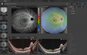
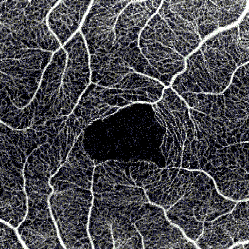
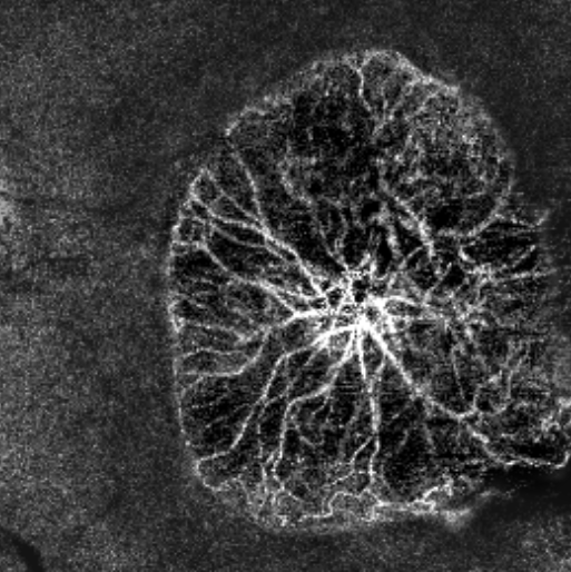
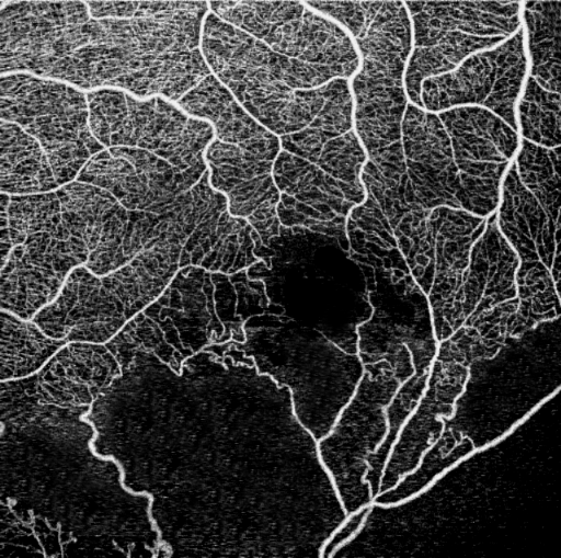
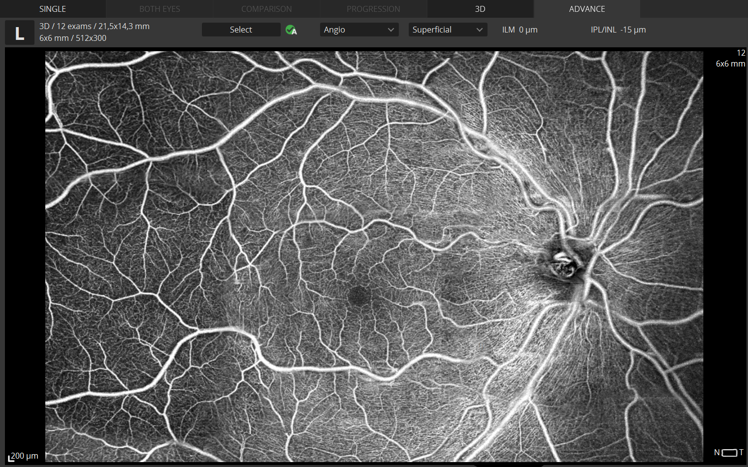
This non-invasive dye free technique allows the visualization of the microvasculature of the retina.
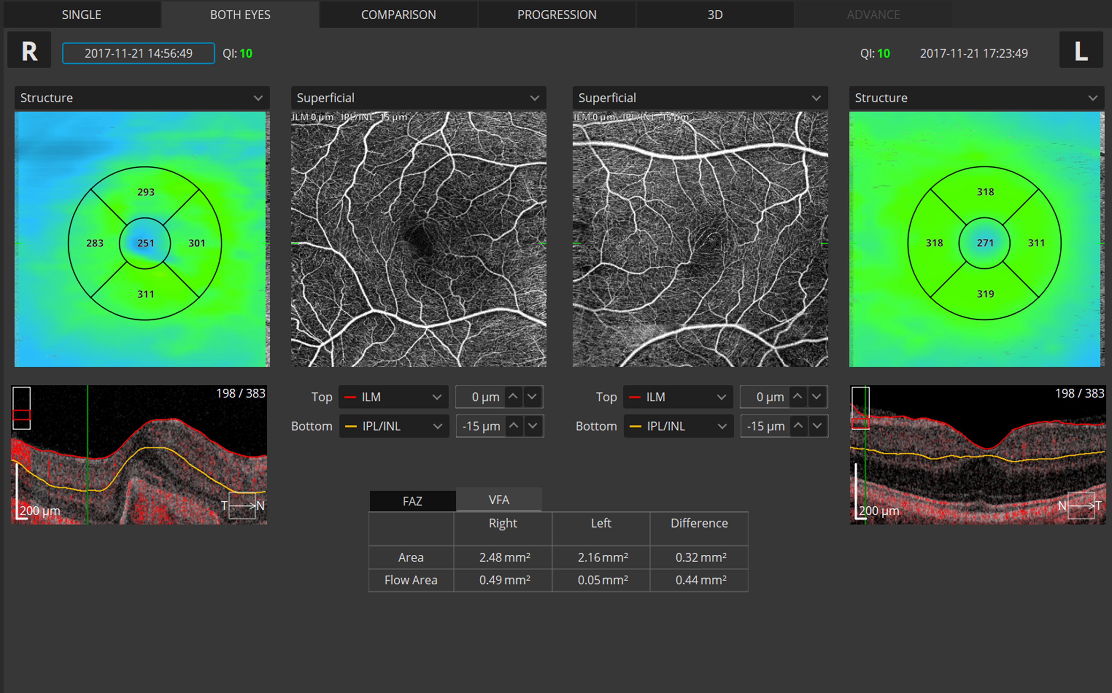
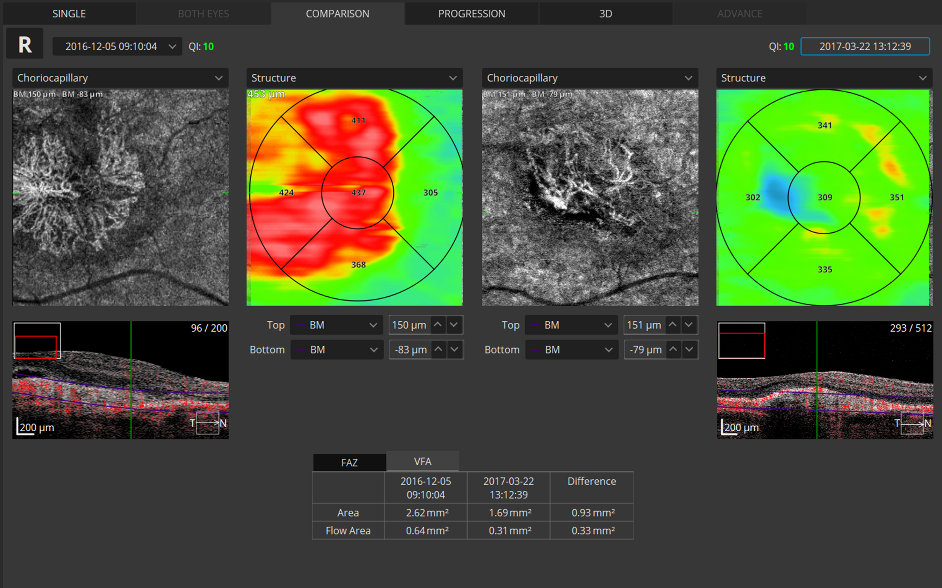
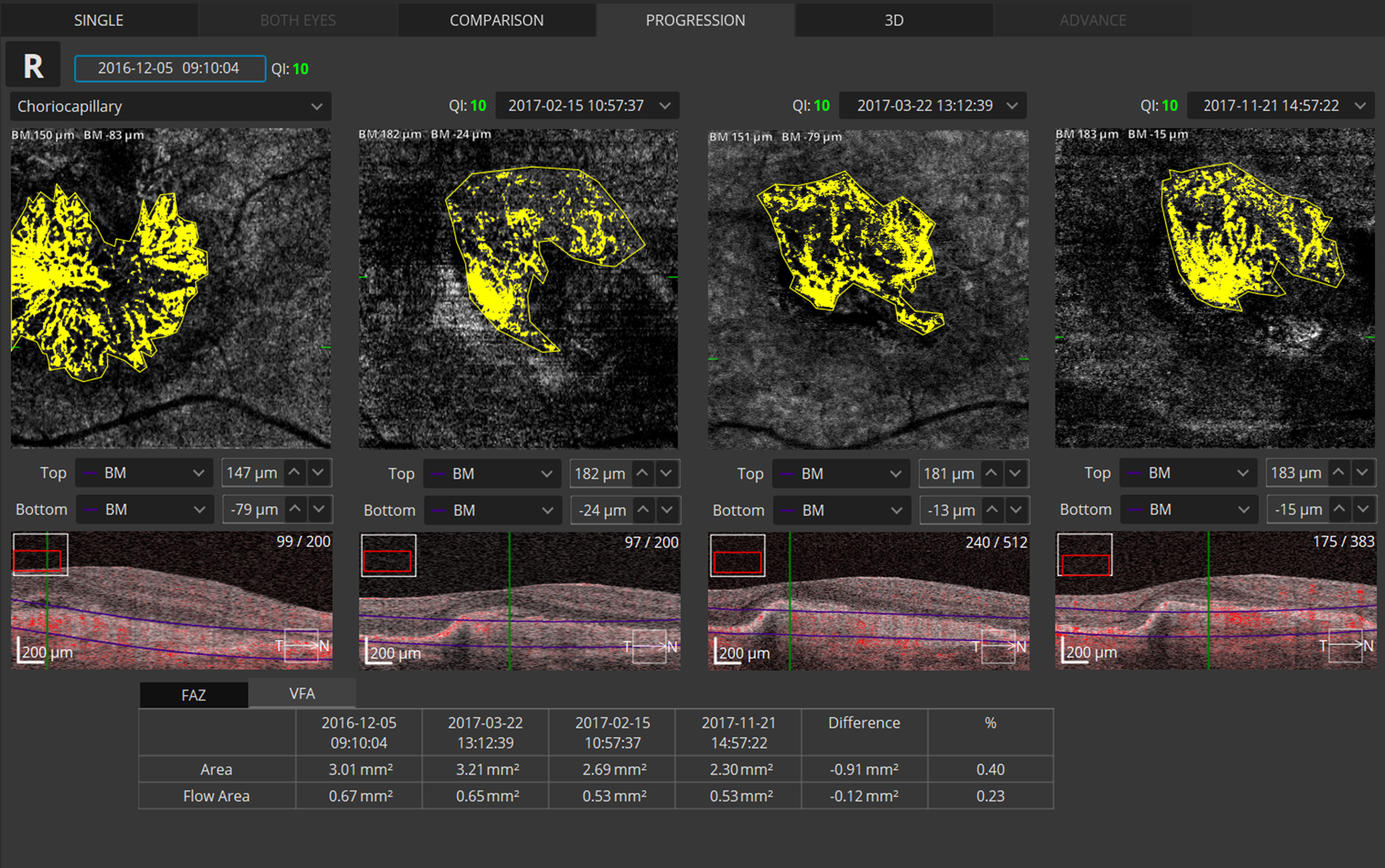
Angiography OCT provides an alternative to the traditional fluorescein method. Although OCT-A will not completely replace FA imaging, it is a quick and non-invasive tool. The software allows clinicians to observe, track and compare changes in the microvasculature of the retina in both eyes.
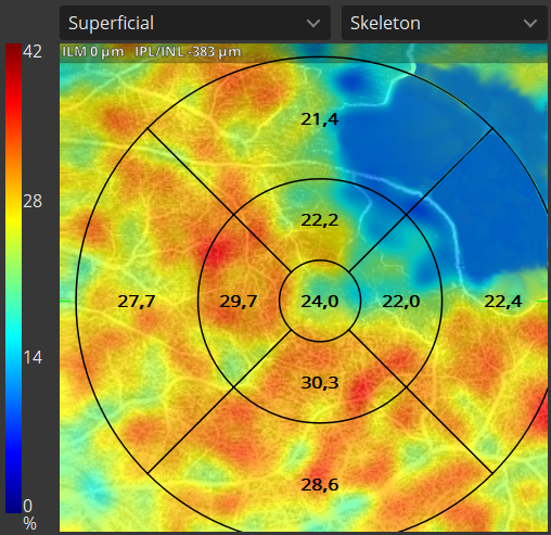
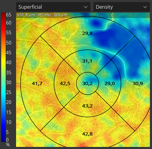
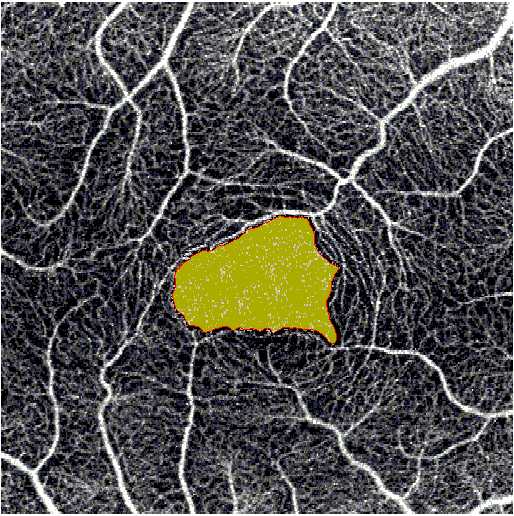
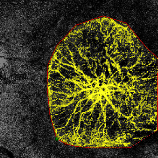
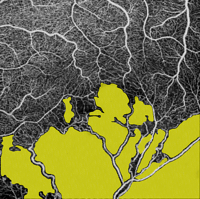
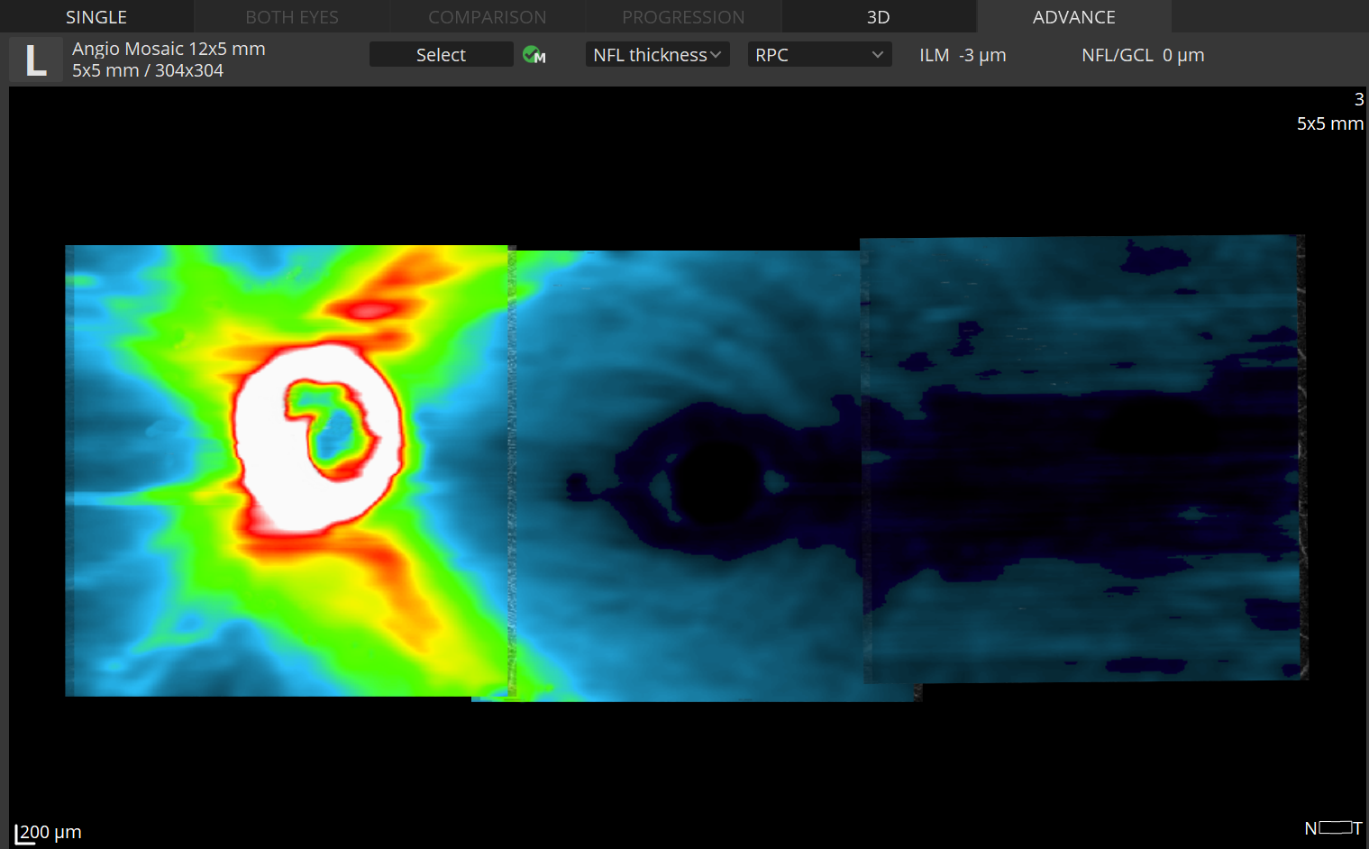
The quantification tool provides quantification of the vasculature in the whole analyzed area together with values in specific zones and sectors. Thanks to the heat map of the analyzed vasculature the evaluation of vascular structure conditions is much faster. The choice of the quantification method increases the sensitivity of analyses for specific deseases.
Available quantification methods:
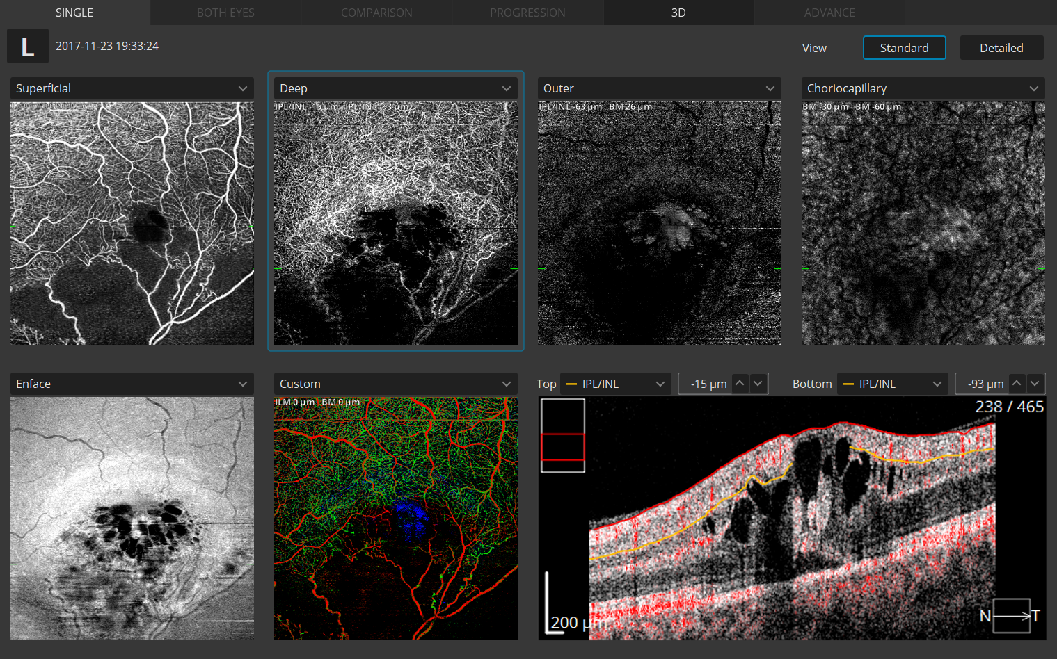
Quantification is available for a specific layer in Angio OCT exam:
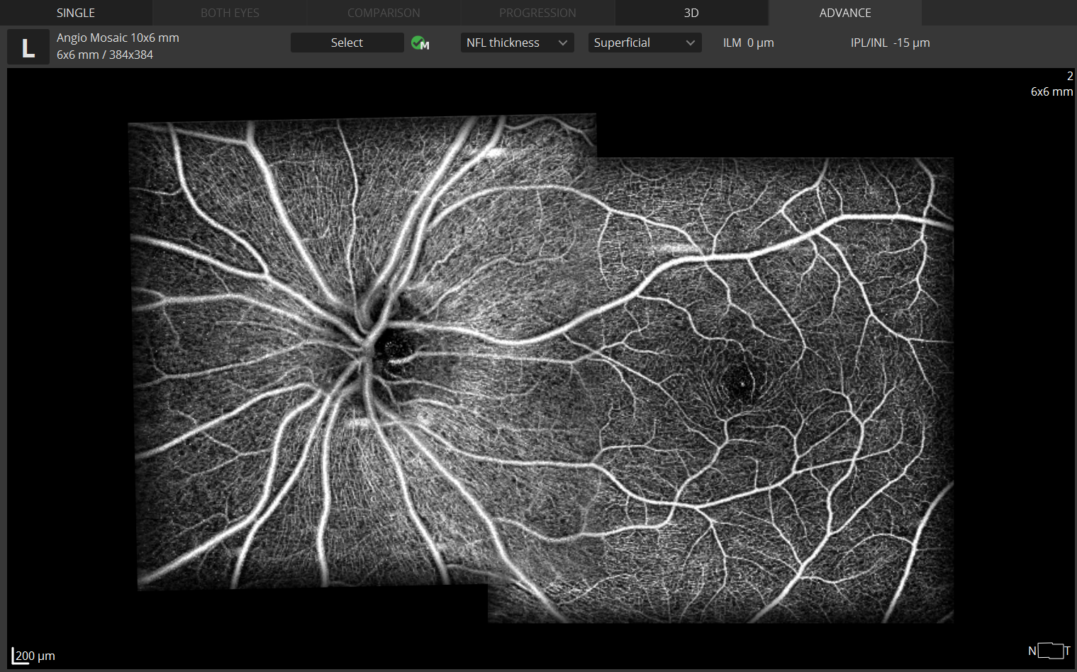
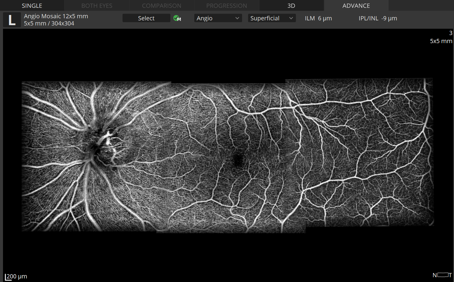
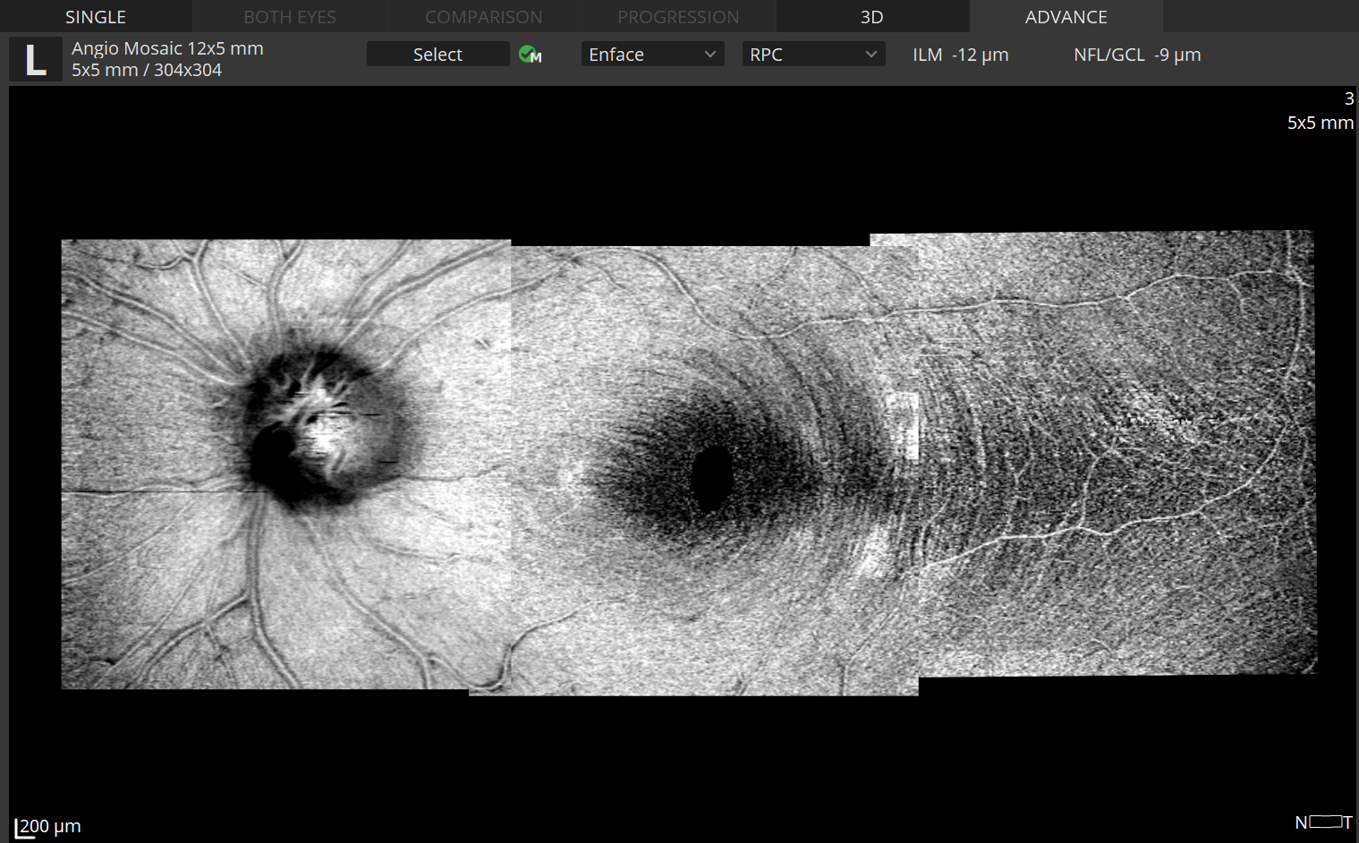
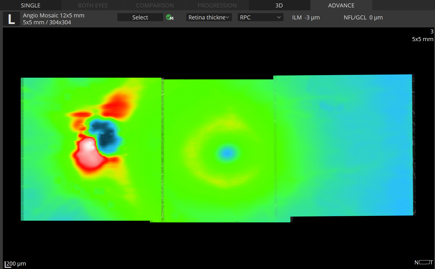
The Angiography mosaic delivers high-detailed images over large field of the retina.
Advanced tab: provides view of any vascular layers, enface view of vascular layers, depth coded and thickness map.
Mosaic modes: 10×6 mm, 12×5 mm, 7×7 mm, 10×10 mm and Manual (up to 12 images).
Manual mode: allows to scan the desired region.Built-in analytics allow to see vascular layers, enface or thick-ness maps.


